Treatment & Management Treatment & Management

For patients requiring cardiac rehabilitation or heart failure management, a comprehensive assessment is fundamental to developing a management plan.  A thorough multidisciplinary assessment helps identify an individual’s risk of deterioration or decompensation; any changes in their conditioning as well as their capacity to self- manage. Essential elements of assessment are described below.

Note factors relevant and potentially contributing to the presentation, including:

  • Onset of recent symptoms (e.g., chest pain, SOB)
  • Cardiac related conditions (e.g., new AF or other rhythm disturbance)
  • Self-management issues (e.g., poor adherence with medications, fluid or salt restriction as appropriate)
  • Medication issues (e.g., non-adherence, recent use of medications that require caution or monitoring)
  • Social issues
  • Recent infection, viral illness, vomiting or diarrhoea
  • Risk of complications or  decompensation

Medical and surgical history should include the main complaint, current problems and any previous illnesses and operations, as summarised below. All health professionals involved in the patient’s care should also be identified.

  • Cardiac conditions: Pre-existing ischaemic heart disease, MI, rheumatic fever, arrhythmia, hypertension, valve conditions, HF
  • Cardiac surgery/intervention: CABG/ valve surgery or percutaneous valvular intervention, percutaneous coronary interventions with or without stenting, electrophysiology studies and interventions
  • Cardiovascular risk factors 
  • Other relevant medical conditions: Anaemia, chronic kidney disease, diabetes, gout, malignancy/chemotherapy, past surgeries/interventions, respiratory disease, relevant childhood illnesses, past surgery, sleep apnoea, thyroid disease. See Co-morbidities and related conditions or further information
  • Family history: E.g., history of MI, cardiomyopathy, sudden cardiac death, congenital heart disease, valvular heart disease, Marfan’s syndrome
  • Implantable devices: Note whether the patient has an implantable cardiac device or if one is being considered. Implantable loop recorders (ILRs), permanent pacemakers (PPMs) and implantable cardioverter defibrillators (ICDs) are used in a variety of circumstances for patients with cardiac disease

The following information should be recorded:

  • Occupation and any relevant tasks associated with occupation, e.g., commercial driver, physical demand
  • Transport and health related restrictions to driving
  • Living situation: Type of residence (e.g., own home, residential care), presence of stairs, availability of assistance (community support services, lives with family or alone, supportive friends or family)
  • Social activity: Is the person socially active or isolated; what are the family relationships and support networks?
  • Literacy: First language and level of health literacy
  • Psychological conditions: Depression and anxiety are common in patients with cardiac disease, with a causal relationship between depression and social isolation and heart disease. Those treated with selective serotonin reuptake inhibitors (SSRIs) may require sodium monitoring as these medications can cause hyponatraemia.

Depression screening should be routine. See Psychosocial issues for patients requiring more in-depth assessment.

For further information see Psychosocial assessment section

The Medication history and review section provides details on undertaking a comprehensive assessment. An initial, general assessment should note:

Current prescribed and over-the-counter (OTC) medications including herbal or natural supplements
Allergies
Medication adherence and how the patient is managing their medications including the use of a dosette box or Webster-pak®
Vaccinations: Check whether flu and pneumococcal vaccinations are up-to-date according to national guidelines. For those with a chronic disease such as HF a yearly flu vaccination is usually advisable
Non-prescription drugs and illicit substances may impact on the person’s physical and psychosocial status and may need referral to support agencies such as drug and alcohol services

See Medication history and review for more information.

A number of pathology and imaging techniques are used to clarify diagnosis, aetiology and response to treatment and will guide the management approach.

See Investigations for cardiac disease for more detail.

For patients with HF, intensive management should be prioritised for those at moderate to high risk of decompensation, or who have newly diagnosed systolic dysfunction. The table below offers guidance on how to judge the risk of decompensation or readmission to hospital for these patients. These factors should be noted during assessment and used to guide the planning and timing of intervention.

Table 1: Assessing patient risk for decompensated heart failure

Low Risk Moderate to High Risk

Class I or II NYHA symptoms of heart failure

Class III or IV NYHA symptoms of heart failure

Knowledge and understanding of condition

Over 65 years

Ability to follow medication and dietary guidelines

Left ventricular ejection fraction <30%

No other admissions for decompensated heart failure within last 6-12 months

Significant renal dysfunction (GFR<60 mL/min/1.73m2)

Adequate social support

Poor understanding of condition including those from a culturally and linguistically diverse background or due to poor cognitive function

Regular contact with GP

Admissions with decompensated HF in previous 6 months


Socially isolated


Multiple co-morbidities and risk factors including depression

  Poly pharmacy/poor adherence with medication 

Adapted from Stewart & Blue 2004 [#stewart-s-blue-l.-2004] and the National Heart Foundation of Australia and the Cardiac Society of Australia and New Zealand guidelines 2011 [#national-heart-foundation-of-australia-and-the-cardiac-society-of-2011]

The patient should be observed to gain an impression about obvious issues. Check to see whether the patient looks unwell, including facial expressions, general movement, obvious respiratory distress, body composition, skin colour, warmth and coolness of the peripheries, odour and voice.

Record vital signs including:       

  • Pulse: Assess rate and rhythm as well as the volume and character
  • Blood pressure (BP): Assess lying as well as standing BP if the patient is symptomatic e.g, experiences dizziness or blackouts on standing

Orthostatic hypotension (or postural hypotension)  is low BP that occurs when standing up from a sitting or lying position and is defined as a drop in SBP by at least 20mmHg or DBP by at least 10mmHg. Orthostatic hypotension can cause dizziness or light headedness, and may even result in fainting. It is often mild, lasting a few seconds to a few minutes after standing. However, long-lasting orthostatic hypotension can be a sign of more-serious problems which requires further investigation.

  • Oxygen saturation: Determine if accompanied by dyspnoea and if this is new or worsening (a 6 minute walk test (6MWT) will identify those requiring routine oxygen saturation via pulse oximetry (SpO2) monitoring)

Additional assessments may be warranted depending on the situation, such as:

  • Wound assessment for those who have recently undergone surgery
  • Skin integrity for diabetics
  • Sleep disturbance is frequently under recognised in patients with CVD and HF. See sleep disturbances for further information

Clinical reasoning should be applied to determine the most appropriate course of action for symptom management. Some symptoms may require urgent medical review, whilst others may be usual for the individual and can be safely self-managed.

For any symptom note:

  • If the onset or change is sudden
  • When it occurs and provoking factors
  • Duration
  • Ameliorating factors
  • Associated symptoms
  • If the patient or carer can safely self-manage

The presence of angina or non-ischaemic chest pain varies significantly from individual to individual. Assessment should therefore identify if medical review or further investigations are warranted and if this is urgently required.

It is important to:

  • Ascertain the patient’s history and nature of chest pain symptoms
    • Record a baseline assessment by asking the patient to describe the quality, location, duration, frequency and intensity of their pain
  • Determine if there are any factors that provoke or relieve the pain. Note if there are any changes or progression of usual symptoms
    ​Does the patient demonstrate an understanding of angina, including ability to:
    • Identify warning signs of a heart attack, i.e., chest pain, upper body and jaw pain and atypical signs such as shortness of breath, nausea, dizziness, clamminess or indigestion
    • Use a short acting nitrate: Question the patient as to whether they carry this medication at all times and are able to describe how to use it properly. Assess their knowledge about short acting nitrate expiry, storage, and indications and contraindications for use
    • Know the number to call emergency if symptoms continue after 10-15 minutes
    • Understand why rapid access to treatment is important
      See Warning signs of heart attack patient fact sheet.

Not all patients experience symptoms of angina. Patients with diabetes are more likely to experience minimal or no symptoms of chest pain, leading to silent ischaemia.

Palpitations are caused by multiple factors and may be benign, but in individuals with cardiac disease, they may be indicative of a cardiac dysrhythmia such as atrial fibrillation (AF). Establish what action the patient takes to manage the symptoms and if there is an action plan. 

There are many cardiac and non-cardiac causes of breathlessness and assessment should aim to identify where further investigations or medical intervention is required.

Record a baseline of symptoms and consider the following:

  • Does breathlessness occur at rest or on exertion and describe the activities?
  • Does it occur only when experiencing chest discomfort?
  • Is orthopnoea (breathlessness when lying flat) or paroxysmal nocturnal dyspnoea (waking at night with breathlessness) present?
  • Ascertain how many pillows are required to sleep without dyspnoea and if there is any change from what is usual, since patients with increasing pulmonary congestion may find it difficult to breathe when lying flat and opt to use additional pillows to assist breathing

For patients with HF, change in weight is an important indicator of fluid status. Compare the patient’s current weight to their dry weight (i.e., when clinically euvolaemic) and evaluate trends in weight over time by giving the patient a daily diary to complete. See Weight and symptom diary.  Note that loss of body mass can mask fluid overload as weight appears to remain stable.  Daily weighing is more likely to detect changes in fluid status as a  rapid weight change of 2 kg (4.5 lbs) over 1-2 days is more suggestive of  fluid overload or dehydration rather  than a change in body mass

Nutrition assessment of patients with HF can be challenging as weight loss may be masked by fluid retention. Use dry weight when assessing weight loss. Despite these difficulties, it is important to routinely screen all HF patients, inclusive of overweight/obese patients, for malnutrition risk using the Malnutrition Screening Tool (MST) [#ferguson-m-capra-s-bauer-j-et-al.-1999] (see table below).

Patients with an MST score of 2 or more should be referred to the Dietitian for further assessment, if available.

Table 2: Malnutrition screening tool

Malnutrition Screening Tool  

1. Has the patient lost (dry) weight recently without trying

Score
No 0
Unsure 2
If yes, how much weight has been lost?  
1 - 5 kg

1

6 - 10 kg  2
11 - 15 kg 3
>15 kg 4
Unsure 2
2. Has the patient been eating poorly because of a decreased appetite?  
No 0
Yes 1
Total Score (>2 refer to dietitian)  

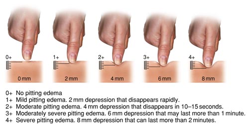
Oedema may be caused by multiple factors such as HF, musculoskeletal disorders, gout, saphenous vein harvesting for CABG (leading to lower limb oedema on the side of the graft); and medications such as calcium channel blockers.

Assess a follows:

  • Observe the presence of oedema and apply gentle pressure on the affected areas, noting the location and whether there is pitting.
  • Rate oedema severity by pressing down firmly to the bone and releasing. Also assess the extent of pitting and how long it takes for the indentation to return to normal.

Figure 1: Oedema rating scale

Oedema rating scale

Table 1: Examinations requiring advanced clinical skills*:

Assessment Rationale for assessment
Abdominal assessment Identifies signs and symptoms of tenderness, liver enlargement and ascites
Jugular Venous Pressure (JVP) Provides a clinical measure of the central venous pressure, fluid status and related cardiac function
Heart sounds Measures heart rate and rhythm, identifies underlying valvular abnormalities (murmurs), and can indicate early signs of heart failure decompensation
Lung sounds Assists in differentiation between cardiac and non-cardiac causes of dyspnoea. Signs of decompensated heart failure include inspiratory crackles in dependent lung segments, and in the case of a pleural effusion, decreased or absent breath sounds on the affected side

*The online resource from the University of California San Diego provides in-depth instructions on performing a cardiovascular examination.

These may be the consequence of the condition itself (e.g., low cardiac output state or cardiac rhythm disturbances), medication related, or due to fluid retention.

Frailty is a state where physiological reserves are reduced, impairing responses to stressors [#clegg-a-young-j-iliffe-s-et-al.-frailty-in-elderly-people.-lancet.-20133819]. It is common in people with cardiovascular disease and is present in almost half of people with heart failure.  Frailty is a strong predictor of outcomes independent of age and NHYA classification [#denfeld-qe-winters-stone-k-mudd-jo-et-al.-the-prevalence-of-frailty-in-hear] and severity of frailty is associated with higher rates of dependency, healthcare utilisation and mortality.

Assessing frailty using a valid and reliable instrument can help guide appropriate and individualised care, which may help to improve quality of life and impact upon prognosis.  Selection of a frailty measure will depend on the clinical purpose and setting.  Estimates of frailty vary by tool and by cut-offs used, and as such results of different tools cannot be directly compared. The most common approaches to measuring frailty include:

Measure Description
Frailty phenotype [#fried-lp-tangen-cm-walston-j-et-al.-2001] Identifies frailty using 5 criteria which include: unintentional weight loss, self reported exhaustion, slow gait speed, low energy expenditure, weak grip strength.
*Clinical frailty scale [#rockwood-k-song-x-macknight-c-et-al] Uses clinical descriptors and pictographs to stratify people into 9 levels of vulnerability.

Edmonton frail scale
[#rolfson-db-majumdar-sr-taher-a-et-al]

10 domains which include cognition, functional performance, mood, functional independence, medication use, social support,  nutrition, health attitudes, continence and quality of life
Frailty index (accumulation of deficits) [#rockwood-k-mitnitski-a] Quantifies total burden of deficits across multiple domains

* http://geriatricresearch.medicine.dal.ca/clinical_frailty_scale.htm

  • Stewart S, Blue L. Improving Outcomes in Chronic Heart Failure: Specialist nurse intervention from research to practice, 2nd Edition. London: BMJ Publishing. 2004.

    stewart-s-blue-l.-2004
  • NHFA CSANZ Heart Failure Guidelines Working Group: Atherton JJ, Sindone A, De Pasquale CG, et al. National Heart Foundation of Australia and Cardiac Society of Australia and New Zealand: Guidelines for the Prevention, Detection, and Management of Heart Failure in Australia 2018. Heart Lung Circ. 2018;27(10):1123-208.

    National-Heart-Foundation-of-Australia-and-the-Cardiac-Society-of-Australia
  • Ferguson M, Capra S, Bauer J, et al. Development of a valid and reliable malnutrition screening tool for adult acute hospital patients. Nutrition 1999;15:458-64.

    ferguson-m-capra-s-bauer-j-et-al.-1999
  • Clegg A, Young J, Iliffe S, et al. Frailty in elderly people. Lancet. 2013;381(9868):752-62

    clegg-a-young-j-iliffe-s-et-al.-frailty-in-elderly-people.-lancet.-20133819
  • Denfeld QE, Winters-Stone K, Mudd JO, et al. The prevalence of frailty in heart failure: A systematic review and meta-analysis. International journal of cardiology. 2017;236:283-9

    denfeld-qe-winters-stone-k-mudd-jo-et-al.-the-prevalence-of-frailty-in-hear
  • Fried LP, Tangen CM, Walston J, et al. Frailty in older adults. J Gerontol A Biol Sci Med Sci 2001;56:M146-157.

    fried-lp-tangen-cm-walston-j-et-al.-2001
  • Rockwood K, Song X, MacKnight C, et al. A global clinical measure of fitness and frailty in elderly people. CMAJ. 2005; 173(5): 489–495.

    rockwood-k-song-x-macknight-c-et-al
  • Rolfson DB, Majumdar SR, Taher A, et al. Development and validation of a new instrument for frailty. Clin Invest Med 2000; 23: 33

    rolfson-db-majumdar-sr-taher-a-et-al
  • Rockwood K, Mitnitski A. Frailty defined by deficit accumulation. J Gerontol A Biol Sci Med Sci. 2007;62(7):722-7

    rockwood-k-mitnitski-a