Exercise Exercise

Assessment of exercise capacity provides valuable information to guide exercise prescription. This includes subjective assessment of an individual’s exercise tolerance, and objective exercise test results, which can be used to calculate exercise intensity based on an equation or algorithm.

All assessments of exercise capacity have strengths and weaknesses in assisting exercise prescription, and many published studies have evaluated their validity in various clinical settings. A summary of the strengths and weaknesses for the most commonly used assessments for exercise capacity is available. [

]

When deciding on the most appropriate exercise test, consider:

  • The primary purpose of the test
  • The workload intensity achieved during the assessment and its implication for risk
  • The clinical risk of the patient and the setting in which testing will be undertaken
  • Whether the assessment will also be used for research or to evaluate your service. If so, strict adherence to testing procedure guidelines will promote validity and reproducibility of results

In clinical settings, submaximal exercise testing is used more commonly than maximal testing as it is easily administrated, less likely to cause adverse events and does not require medical supervision and ECG monitoring.

Submaximal tests are defined by an age-predicted HR <85% calculated by the Gelish formula (maximum HR = 206.9 - 0.67 x age) or a RPE <15 (Borg scale of 6-20).

TIP: Most submaximal, non-diagnostic tests are performed while the patient is taking their normal medication. HR-based calculations are complicated by medications that alter HR response during exercise (e.g., such as beta-blockers). Therefore, it is important to document all medications at the time of assessment and consider cardiac medications that impact on exercise response

HR response may not be a reliable guide to exercise prescription so it is useful to also use the Rating of perceived exertion (RPE) - Borg scale.

The cardiopulmonary exercise test (CPET) is an example of a maximal exercise test. Maximal tests are characterised by HRs greater than 85% of age-predicted values off medication (maximum HR = 206.9 - 0.67 x age) or a rating of perceived exertion >15 (6-20 Borg scale; RPE).

Maximal exercise testing allows for:

  • More precise exercise prescription than submaximal testing and greater reproducibility for follow up assessment
  • Identification of cardiovascular compromise at higher levels of exercise and provision of an individualised maximum HR on which submaximal prescription can be based
  • Gradation of exercise until the patient experiences signs/symptoms of cardiovascular compromise or reaches volitional exhaustion, or some other limiting symptom

A CPET is similar to an exercise stress test, with the addition of ventilatory gas analysis to determine peak oxygen consumption (VO2 peak). Oxygen consumption is determined by central (heart) and peripheral (muscle) factors that respectively influence the body’s capacity to pump and utilise oxygenated blood.

A CPET is commonly used to determine prognosis in patients with HF and to stratify patients for cardiac transplantation.

  • A VO2 peak <14 mL/kg/min (or <12 mL/kg/min in those tolerating optimal beta-blocker doses) identifies patients with poor 1-year survival who are likely to benefit from cardiac transplantation
  • In patients under 50 years of age, VO2 peak <50% of the value predicted for age supports consideration for transplant listing
  • If the CPET is deemed submaximal (respiratory exchange ratio <1.05), a ventilation equivalent of carbon dioxide (VE/VCO2) slope of >35 may also help determine transplant listing

TIP: Maximal exercise tests have a risk of adverse events, so they require a medically supported environment with 12-lead ECG and BP monitoring.

The six-minute walk test (6MWT) is an internally (self-paced), walking test well suited to a hospital or community setting. An abundance of resources relate to its use in many clinical settings, most of which are adapted from the European Respiratory Society/American Thoracic Society technical standard [

].

Useful resources include:

The test is undertaken using strict guidelines to ensure standardisation. A 30-metre track is recommended where space is available and standardised instructions are given. Heart rate, blood pressure, oxygen saturation, RPE and the presence of any symptoms should be recorded.

Perceived exertion is particularly important for patients taking beta-blockers as the HR response will not accurately reflect exercise intensity. The test should be terminated if the patient's HR exceeds the pre-determined limit or if concerning symptoms develop.

A second test is commonly recommended to account for a learning effect. This is particularly important for patients whose baseline 6MWT distance is >300m. For patients with more severe classes of heart disease, 6MWT results correlate well with VO2 peak, and only one 6MWT is usually required. For information about prescribing walking programs based upon 6MWT results, see Walking prescription 6MWT as an outcome measure.

Change in 6MWT distance can be measured in several ways. The most common include:

  • Absolute change (post-program distance - pre-program distance)
    The minimum important distance (MID) is 25m in patients with coronary artery disease (CAD) and 36m for patients with chronic HF.
  • Percentage change
    This may be a more relevant measure for frail patients whose baseline distance is very short (e.g., <100m), and can be calculated as follows:
    % change = (post-program distance ‒ pre-program distance) ÷ pre-program distance x 100

Reference equations which adjust for variables such as height, weight, age and gender, are used by some clinicians to predict clinical progress. An example of these is listed below.

Males : 6MWD = 867 – (5.71 x age, yrs) + (1.03 x ht, cm)
Females : 6MWD = 525 – (2.86 x age, yrs) + (2.71 x ht, cm) – (6.22 x BMI)

Another submaximal exercise test is the Incremental shuttle walk test (ISWT).  Whilst this externally paced test is commonly used in pulmonary rehabilitation programs, it is not frequently used in cardiac rehabilitation. The ISWT may be beneficial for patients with few symptoms who may reach a ceiling effect in the 6MWT.

The sit to stand test (STST) is easily performed, requires minimum time and equipment and closely resembles daily functional activities.  As such, it is a valuable outcome measure in acute and subacute care settings. In recent years, it has also become popular for home-based and virtual cardiac and heart failure rehabilitation programs. 

Several varieties of the STST are available, the most common of which are the 5 times sit to stand (5STST), the 30 second sit to stand (STST-30) and the 60 second sit to stand test (STST-60). Whilst the STST-60 has been shown to elicit similar physiological responses to the 6MWT, the 5STST is less demanding and thought to correlate more closely with lower limb strength.

Test procedure

  • The test commences with the patient sitting in a chair of standard height (46-48 cm), with arms crossed across the chest
  • Upon command, the patient stands to a full standing position and returns to sitting
  • For the 5STST, the time taken to complete 5 cycles of sitting to standing is recorded
  • For the STST-30 and STST-60, the number of complete stands completed during the respective time period is recorded.
  • If the patient is unable to stand without using their arms for assistance, they are scored 0.
  • Only 1 test is required due to high levels of reliability

The minimum important clinical difference has not yet been reported for cardiac populations, however for other populations is:

  • 5STST – 2.3 seconds
  • STST-30 – ≥ 2 repetitions
  • STST-60 – 3 repetitions

In addition to the sit to stand test, other functional testing may be relevant for some individuals to assist exercise prescription and assessment of program outcomes. Some examples of other tests commonly used in cardiac and HF rehabilitation include:

  • Muscle strength testing 
  • Timed up and go tests (TUGT)
  • 10 metre walk speed
  • Balance and flexibility tests

Muscle strength testing is often included in the assessment to:

  • Determine the appropriate load for resistance training
  • Provide a global measure of muscle strength as a pre- and post-program outcome measure
  • Assess strength deficits in specific muscles, thereby guiding exercise prescription

In patients with cardiac conditions, muscle strength can be assessed in several ways including [

] :

  • 1RM method - the maximum weight lifted in one full range of motion. This method can be used for any major muscle group.
  • Estimated 1 RM assessments - may be conducted by recording the highest weight an individual can lift to failure (eg. 3 repetitions) or a graded approach such as 10-15 RM.
  • Hand-held dynamometry
  • Five times sit to stand test

Grip strength

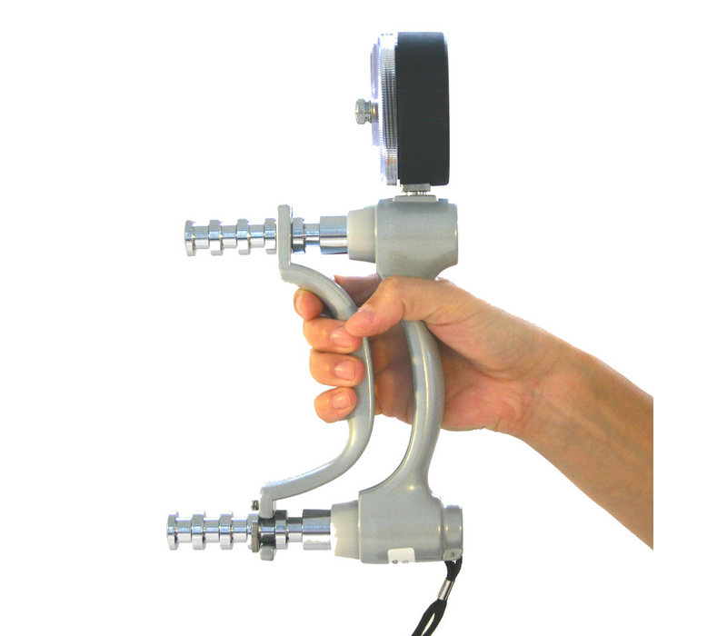
Grip strength is measured quantitatively using a hand-held dynamometer (see example below).

Hydraulic_hand_dynamometer

Reproduced with permission from Optomo

Weak grip strength is associated with falls, disability, impaired health-related quality of life, prolonged length of stay in hospital and increased mortality.

The following are important to ensure accuracy with repeat testing:

  • Body position:  seated, shoulders adducted and neutrally rotated
  • The upper arm should be by the side with the elbow flexed at 90°, the forearm in neutral and wrist between 0-30° of extension
  • Adjust the device for hand size
  • Record hand dominance
  • Record the maximum or the mean of three trials in each hand

Assessment of other muscle groups

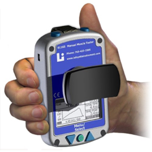
Quadriceps and hamstring strength may be measured using a hand-held dynamometer (e.g., Lafayette Manual Muscle Test System – see below) using the protocol described by O'Shea et al. [

]

Lafayette Manual Muscle Testing System

Lafayette_Maunal_Muscle_Test_System

Reproduced with permission from Lafayette Instrument

  • 1

    A Clinical Guide for Assessment and Prescription of Exercise and Physical Activity in Cardiac Rehabilitation. A CSANZ Position Statement. Heart Lung Circ. 2023.


    a-clinical-guide-for-assessment-and-prescription-of-exercise-and-physical-a
  • 2

    Holland AE, Spruit MA, Troosters T, et al. An official European Respiratory Society/American Thoracic Society technical standard: field walking tests in chronic respiratory disease. Eur Respir J. 2014;44:1428-1446.

    holland-ae-spruit-ma-troosters-t-et-al.-2014
  • 3

    O'Shea SD, Taylor NF, Paratz JD. Measuring muscle strength for people with chronic obstructive pulmonary disease: Retest reliability of hand held dynamometry. Arch Phys Med Rehabil. 2007;88: 32-36.

    oshea-sd-taylor-nf-paratz-jd.-2007